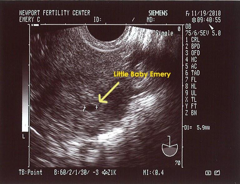

Sometimes transabdominal ultrasound scans are true to the same maternal side. The debate among pregnant women who discuss the Ramzi Theory is questioning whether transvaginal ultrasounds appear to be on the “same side” as pictured on the scan because abdominal scans are “flipped” (or mirror image)īecause of this, The Gender Experts rule-of-thumb is : Not all scans are created equal for the Ramzi Theory. The wand will not be painful, although you will feel some pressure and maybe some mild discomfort. A specially designed ultrasound wand is inserted into the vagina, to give a clearer and more detailed view the uterus.

Both abdominal and transvaginal scans can be used when predicting with the Ramzi Method. 6 week transvaginal ultrasound An ultrasound during early pregnancy is usually a transvaginal ultrasound. Internal scans are often used early in the pregnancy to assist the doctor or technician in obtaining a high-quality view.īecause of the nature of a transvaginal ultrasound, your technician will be able to determine both the left and right sides of your uterus, and therefore can help you find where (which side) the placenta is located. These photos can capture images of the vagina, cervix, uterus, fallopian tubes, and even the ovaries. So essentially – a square-shaped little skull for a boy, and round itty bitty head for a girl.Unlike an abdominal ultrasound, a transvaginal-sometimes called an endovaginal-scan is done when the ultrasound wand is inserted 2 or 3 inches inside the vaginal canal in order to take photos from inside. For boys you’re looking for a more square skull and jaw because men have lower and more sloping foreheads.įor a girl, the top of the head is rounder and tapers at the top and the cheekbones are less pronounced. This method of gender prediction is based on the shape of your unborn babe’s skull. This method is based on the genital tubercle or the ‘nub’ that babies have between their legs between 11 and 13 weeks. If the nub points up more than 30 per cent, it’s a boy. The theory goes that if the nub is pointing up less than 30 per cent from the spine, it’s a girl. There are entire forums dedicated to the gender prediction nub theory, where you can ask thousands of people what they think you’re having based on your ultrasound and the direction of bub’s genital ‘nub’. However, studies since have disproved the theory (but it’s still fun to see if it’s accurate for you). On the right side of the uterus for a boy and on the left side of the uterus, for a girl. According to a research paper more than 5000 women had an ultrasound at six weeks to see where their placenta was forming, and they had a follow-up ultrasound at 18 to 20 weeks to find out their baby’s gender.Īccording to the study, the side that the placenta had formed on was correct in predicting the baby’s sex around 99 per cent of the time. Placenta positionĪlso known as the Ramzi theory, this method of gender prediction is all based on the position of the placenta. The labia lips on either side of the clitoris sort of looks like a little hamburger, or three lines. For a boy, you’ll see a ‘turtle’ – the tip of the penis peeping ever so slightly from behind the testicles. A ‘hamburger’ sign will usually mean it’s a girl. It’s pretty much what an ultrasound tech is looking for if they’re checking bub’s gender. If there’s a clear ‘between the legs’ image, you may be able to spot either a hamburger or turtle (really!). So, here are four ways to ‘predict’ baby’s gender from an early ultrasound. While mums-to-be now have the option of having non-invasive Prenatal Testing at 10 weeks, which is 99 per cent accurate in predicting gender, for those opting for the traditional 12-week ultrasound, there’s a bit of fun to be had in trying to guess whether there’s a boy or girl on board. In fact, there’s one ‘scientific’ method that claims a baby’s gender can be predicted as early as six-weeks, using an ultrasound.

And apparently there are clues hidden in that first ultrasound image that may just tell you if you’re having a boy or a girl.

The grainy, black and white 12-week ultrasound that makes it all seem so real.


 0 kommentar(er)
